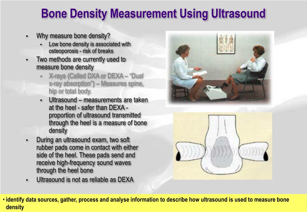
Describe How Ultrasound Is Used to Measure Bone Density
A bone density test is used to measure bone mineral content and density. It is portable inexpensive and involves no radiation.

Quantitative Ultrasound Method A Ultrasound Beam Through A Bone Download Scientific Diagram
For various reasons the DEXA scan is considered the gold standard or most accurate test.
. These tests are often used for screening purposes and can help identify people who might benefit from follow-up bone density testing at the hip and lumbar spine. The bone density measurement using ultrasound technique is another way to assess the bone mineral density BMD with the objective of evaluation of patients at risk for osteopenia and osteoporosis. The use of ultrasound is a highly reproducible measure to assess bone characteristics in a population of pregnant adolescent and young adult women and its summary measure of bone mass is correlated with ethnic as well as body size characteristics.
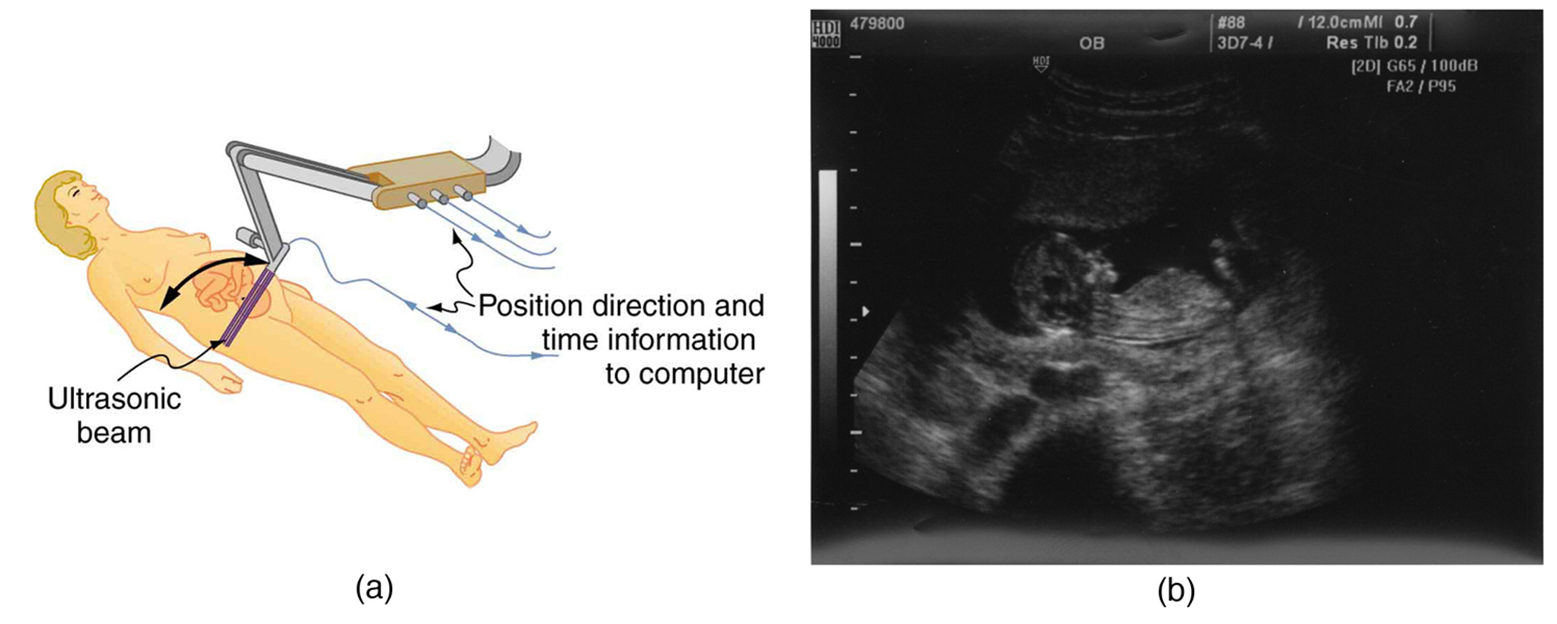
This disparity in test results inspired me to dig a little deeper into the Echolight REMS research. An x-ray generator is located below the patient and an imaging device or detector is positioned above. Over the last decade significant progress has been made in developing technologies to measure bone quality.
This chapter discusses the purpose principle of operation specifications and applications of bone density measurement using ultrasound method. Although measurements of aBMD by DXA were low replicating results from our and others previous studies in this patient population 2 3 23 we found a paradoxically higher SOS at both the radius and the tibia in young women with AN. The sound waves are reflected back to the transducer by boundaries between tissues in the path of the beam eg.
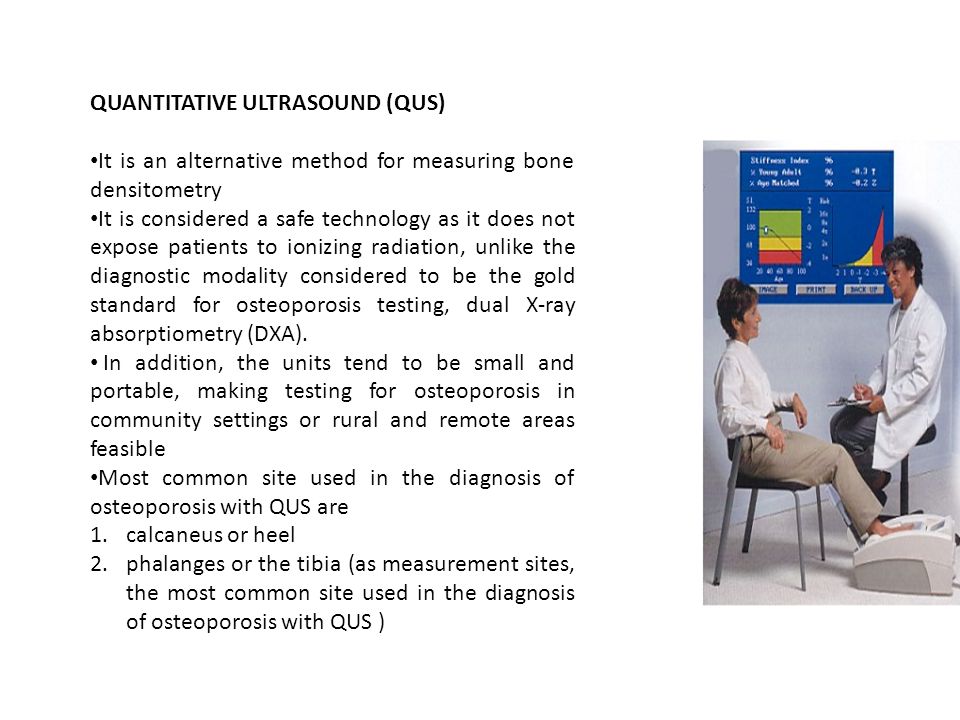
Identify data sources gather process and analyse information to describe how ultrasound is used to measure bone density For the last decade ultrasound has been used to assess bone density to screen for osteoporosis. The bones that are most commonly tested are in the spine hip and sometimes the forearm. When used in an ultrasound scanner the transducer sends out a beam of sound waves into the body.
The boundary between fluid and soft tissue or tissue and bone. Although some have said that ultrasound measures the quality of bone more careful studies suggest that it mainly measures the bone mass. Whereas bone mass is measured with bone mineral density quantification of bone quality is more complex and involves bone architecture texture and mechanical parameters.
Measurement of bone mineral status may be a useful tool in identifying the children who could be exposed to an increased risk of osteoporosis in adulthood. To evaluated bone density using both dualenergy Xray absorptiometry and quantitative ultrasound techniques and examined the changes in body composition in patients. Ultrasound may be a little cumbersome and expensive for measuring density.
Looking at their published articles and talking to individuals. In our system we used Net Delay Time NDT value pressure detection and. Adding an ultrasound measurement to a DEXA does not improve the prediction of fractures.
The speed of sound in the bone is measured. The machine works by measuring how sound waves move through the bone in the heel. For these reasons ultrasound is normally only used as a bone density test when DXA scans are not available.
QUS techniques are safe easy to use radiation-free and devices are portable so that they are particularly indicated to assess bone mineral status in children. The broadband ultrasound attenuation BUA a nd the sp eed of sound SOS are usually measured to evaluate the. Ultrasound could lead to increased screenings for osteoporosis.
These include novel low-cost modalities such as trabecular bone score measured on dual. You should be able to get sensors directly designed for pH and moisture and of course temperature. The test uses X-rays to measure how many grams of calcium and other bone minerals are packed into a segment of bone.
There are limited guidelines for interpreting ultrasound tests to predict fracture risk or diagnose osteoporosis. Indeed DEXA only measures bone density which accounts for about 70 of bone strength. In the last years quantitative ultrasound QUS methods have been developed to assess bone mineral status in some peripheral skeletal sites such as calcaneus phalanges of the hand and tibia.
As it turned out however the two bone density measuring devices delivered quite different results. These results are combined to give the Quantitative Ultrasound Index QUI or Stiffness. To assess the spine the patients legs are supported on a padded box to flatten the pelvis and lower spine.
However as previous stud ies. In the central DXA examination which measures bone density of the hip and spine the patient lies on a padded table. A DXA scan is preferred over ultrasound for measuring bone density.
The machine works by measuring how sound waves move through the bone. In this study we have evaluated quantitative ultrasound as a tool to measure bone density in adolescents and young women with AN. Ultrasound machines became popular as a screening tool for osteoporosis because they are portable not too expensive involve no radiation and do not require a licensed technician to operate.
This technique has not been used as often because there is not as much data about effects of medications. However as previous studies showed that the BUA is sensitive to repositioning and soft tissue the reproducibility of measurement will be difficult to achieve. The new ultrasound device reported substantially lower readings than the DEXA.
A bone density test determines if you have osteoporosis a disorder characterized by bones that are more fragile and more likely to break. It may be done using X-rays dual-energy X-ray absorptiometry DEXA or DXA or a special CT scan that uses computer software to determine bone density of the hip or spine. The broadband ultrasound attenuation BUA and the speed of sound SOS are usually measured to evaluate the bone density.
Bone mineral density BMD based on an ultrasound measurement of the calcaneus. It does not actually measure bone mineral density but speed of sound SOS in metressecond and broadband ultrasound attenuation BUA in decibelsmegahertz. If your pans for raising the dough are standardized then you could just use a webcam to measure an outline and infer a volume from that.
To assess the hip the patients foot is placed in a. 9797487 Indexed for MEDLINE MeSH terms. Ultrasound measurements of bone density are taken with the heel bone as it is flat and a weight bearing site.
Indeed this method offers several advantages over DEXA. Peripheral bone density tests measure bone density in the lower arm wrist finger or heel. It is more simple to use than DEXA the test is rapid and radiation free and it provides information about the quality of bone.
Quantitative Ultrasound Qus What Is Ultrasound Sound Waves Of Extremely High Frequency Inaudible To The Human Ear Ultrasound Can Be Used To Examine Ppt Download
Ultrasound Physics
Ppt Medical Physics Ultrasound Powerpoint Presentation Free Download Id 5588587
No comments for "Describe How Ultrasound Is Used to Measure Bone Density"
Post a Comment